

What if the reason you’re exhausted all the time is hiding in a blood test you never asked for?
You might not have pain, swelling, or a fever—just this low-level tiredness sleep won’t fix.
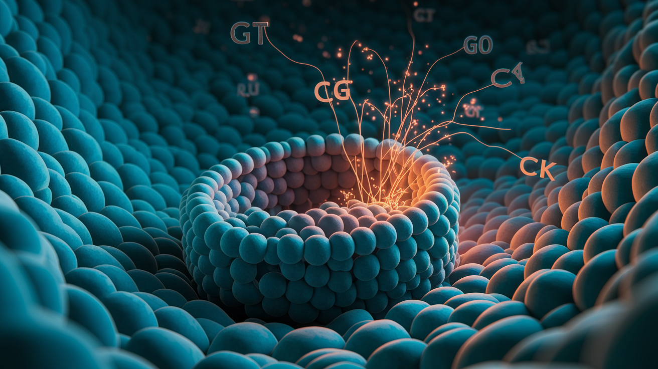
This post points to the key inflammation markers—CRP, ferritin, IL-6, GGT and a few others—that often explain an immune-driven energy drain.
I’ll show how these tests link to your cells’ tiny batteries (mitochondria), iron use, and oxidative stress.
And I’ll give the small next steps you can ask your clinician to run and interpret.
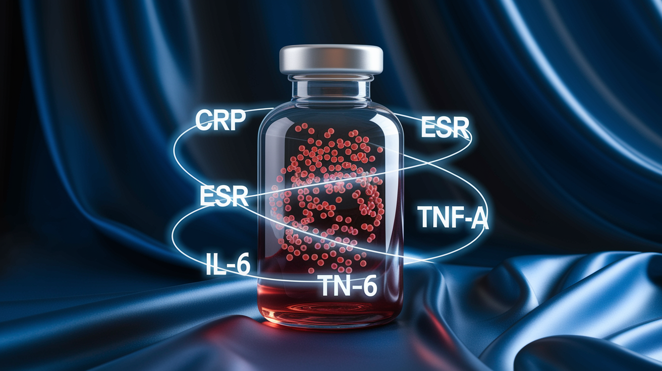
Key Chronic Inflammation Markers That Help Explain Persistent Fatigue
If you’ve been dragging for weeks (or months) without any clear reason, there’s a decent chance the answer’s already hiding in a blood test you just haven’t asked for yet. Chronic inflammation doesn’t always show up the way you’d expect. No pain. No swelling. No fever. Just this low-grade exhaustion that won’t budge no matter how much you sleep.
Some blood markers can pick up on that quiet inflammatory activity before it turns into something you’d actually get diagnosed with. They track how your immune system’s responding, where oxidative stress is building up, and whether inflammatory molecules are messing with your cells’ ability to make energy. When inflammation sticks around too long, it quietly breaks down the systems your body uses to keep you going. That’s where the fatigue comes from, and it’s the kind conventional testing tends to miss.
Here are the markers that matter most when you’re trying to figure out if inflammation’s draining your tank:
- C-reactive protein (CRP) – shows general inflammation across your system
- Erythrocyte sedimentation rate (ESR) – old-school screening test still used for inflammatory conditions
- Interleukin-6 (IL-6) – pro-inflammatory molecule tied to aging and chronic fatigue
- Tumor necrosis factor-alpha (TNF-α) – immune signal that spikes in autoimmune and metabolic issues
- Ferritin – iron storage protein that goes up during inflammation (and can hide real iron problems)
- Gamma-glutamyl transferase (GGT) – tells you about oxidative stress and glutathione use
- Creatine kinase (CK) – points to muscle or metabolic strain
- Vitamin D (25-OH) – low levels connect to more inflammation and mitochondrial trouble
Understanding How Inflammation Markers Connect to Fatigue Physiology
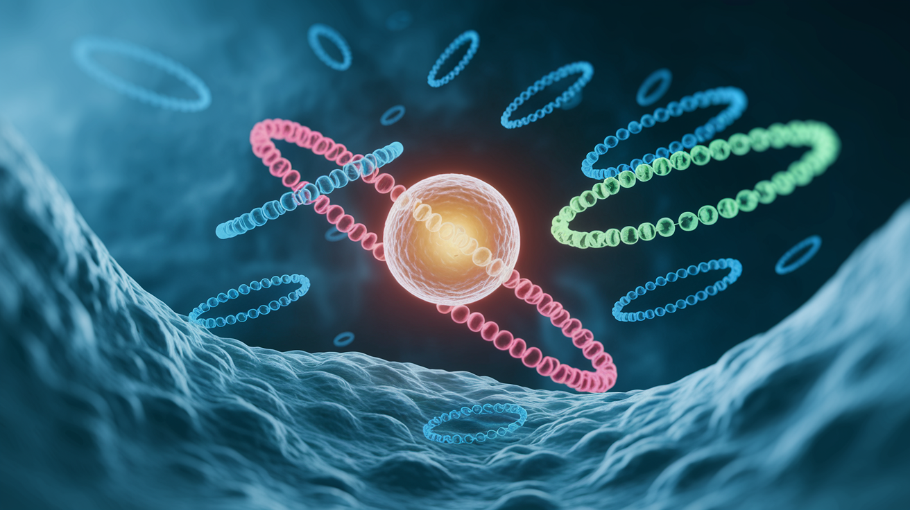
Chronic inflammation doesn’t just make you achy. It actively stops your cells from producing energy the way they should. When your immune system stays turned on for too long, it floods your body with signaling molecules called cytokines. These cytokines basically tell your system to focus on defense instead of normal metabolism. Less energy gets allocated to daily life.
At the cell level, ongoing inflammation damages your mitochondria (the parts that make ATP, which is how your body powers everything). It also cranks up oxidative stress, which you can think of as rust slowly building up inside your cells. Over time, this creates something researchers call “inflammaging,” where your body ages faster biologically and fatigue becomes your new baseline.
Here’s the step-by-step breakdown of how inflammation drains you:
- Cytokine interference – IL-6 and TNF-α make your mitochondria less efficient and signal your brain to slow down (this is why rest feels mandatory).
- Oxidative stress pileup – inflammatory processes create reactive oxygen species that damage your cellular machinery and burn through your antioxidant reserves.
- Iron gets locked away – inflammation triggers your body to sequester iron in storage proteins like ferritin, making it unavailable for oxygen transport or energy production.
- Insulin resistance kicks in – chronic inflammation messes with insulin signaling, so glucose can’t get into your cells even when blood sugar’s normal. Your cells starve for fuel.
Core Blood Tests Used to Identify Chronic Inflammation Fatigue Markers
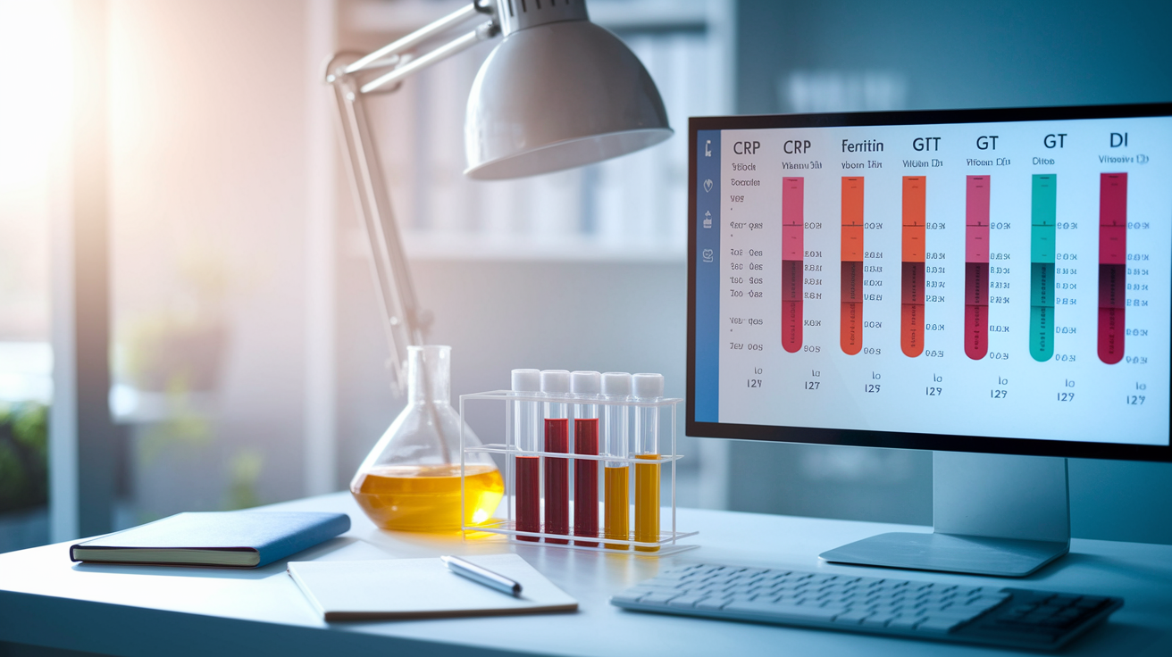
Most inflammation markers tied to fatigue show up on routine lab panels. But you usually have to ask for them by name. A comprehensive metabolic panel (CMP), complete blood count (CBC), liver panel, iron studies, and vitamin D test cover most of what you’d want. Some clinics offer cytokine panels or multi-marker inflammation profiles that check 15 to 22 biomarkers at once.
Context is everything. One elevated marker could just mean you had a bad week, you’re fighting off a cold, or you didn’t hydrate well before the test. But when multiple markers stay elevated over time, you’re seeing the real pattern driving your fatigue. Like if your ferritin’s high but your transferrin saturation’s low and your GGT’s creeping up, that tells you inflammation’s blocking your ability to use stored iron. On paper, your iron looks fine. In reality, your body can’t access it.
You’ll need to fast for accurate insulin and lipid readings. Timing matters for cortisol or hormone tests. Most inflammation markers stay stable throughout the day, but checking the same markers every few months helps you tell the difference between a temporary spike and a chronic issue.
| Marker | What It Indicates | Concern Threshold |
|---|---|---|
| CRP | General systemic inflammation | > 3.0 mg/L (mild); > 10 mg/L (significant) |
| Ferritin | Iron storage and inflammatory activity | < 40 ng/mL (fatigue risk); > 150 ng/mL (inflammation suspect) |
| GGT | Oxidative stress and toxin load | > 18 IU/L |
| Magnesium | Functional deficiency affecting ATP synthesis | < 2.2 mg/dL |
| Vitamin D (25-OH) | Immune regulation and mitochondrial function | < 50 ng/mL |
Cytokines and Immune Signaling Markers Related to Chronic Fatigue
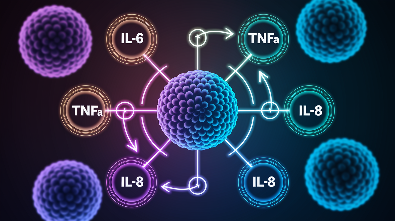
Cytokines are the chemical messengers your immune cells use to organize a defense. When inflammation goes chronic, these messengers don’t turn off. They stay elevated and start interfering with normal metabolic processes. Four stand out when it comes to fatigue and low energy.
IL-6
Interleukin-6 is probably the most studied pro-inflammatory cytokine. It rises naturally as you age and stays high in conditions like obesity, autoimmune disease, and post-viral fatigue (yes, including long COVID). IL-6 directly tells your brain to slow down and conserve energy. That’s why elevated levels often match up with brain fog and the feeling that even basic tasks are too much.
TNF-α
Tumor necrosis factor-alpha got its name because of its role fighting tumors. But now we know it’s a major driver of chronic inflammation and metabolic dysfunction. High TNF-α reduces insulin sensitivity, interferes with thyroid hormone conversion, and promotes muscle breakdown. You’ll see it elevated in inflammatory bowel disease, rheumatoid arthritis, and metabolic syndrome.
IL-1β
Interleukin-1 beta gets released by senescent cells (old cells that won’t die but also won’t function right). These cells leak out a toxic mix of inflammatory molecules called SASP factors, which damage nearby tissues and speed up aging. IL-1β is a key part of that mix. It’s tied to the deep, heavy fatigue you see in chronic fatigue syndrome and fibromyalgia.
IL-8
Interleukin-8 is a chemokine that recruits immune cells to inflamed areas. When it stays elevated throughout your system, it means immune activation and tissue stress aren’t resolving. IL-8 is part of the SASP profile and often shows up alongside other senescent-cell markers in people dealing with age-related energy decline.
Ferritin, Iron-Utilization Patterns, and Inflammation-Driven Fatigue

Ferritin’s tricky because it does two jobs at once. It stores iron, and it acts as an alarm signal during inflammation. When your body senses inflammation, it raises ferritin to lock iron away from invading bugs. Problem is, that also locks it away from your own cells. You end up with a functional iron deficiency even when ferritin looks totally normal or even high.
The sweet spot for energy is 45 to 80 ng/mL. Below 40 ng/mL and fatigue becomes much more likely, even if your hemoglobin’s fine (so technically you’re not anemic). Above 150 ng/mL usually points to inflammation or liver stress, not healthy iron storage. If ferritin’s high but transferrin saturation’s low, that’s your clue inflammation’s blocking iron use.
Common mistakes people make with ferritin:
- Seeing a result of 60 ng/mL, calling it “normal,” and ignoring the fatigue
- Treating high ferritin by restricting iron when inflammation’s the real issue
- Missing functional iron deficiency when ferritin’s sitting at the low end of “normal”
- Not checking transferrin saturation and hemoglobin when interpreting ferritin
- Forgetting to retest ferritin after addressing inflammation to see what the real iron status is
Oxidative Stress and Mitochondrial Function Markers That Contribute to Fatigue
Oxidative stress happens when your cells generate more reactive oxygen species than they can neutralize with antioxidants. This damages mitochondria, the tiny energy factories in every cell. When mitochondria are stressed or damaged, they make less ATP. You feel it as constant low energy, muscle weakness, and sluggish recovery from any kind of effort.
Gamma-glutamyl transferase (GGT) is one of the easiest ways to check oxidative stress. It shows up on most liver panels. Values above 18 IU/L suggest you’re burning through glutathione (your master antioxidant) faster than you can replace it. Common culprits include environmental toxins, alcohol, heavily processed diets, and chronic inflammation. Elevated GGT plus fatigue usually means mitochondrial oxidative stress is draining your energy at the cellular level.
Creatine kinase (CK) is another useful one. It’s an enzyme that gets released when muscle tissue is stressed or breaking down. The reference range is often 65 to 135 U/L. Above 135 U/L can mean metabolic stress or overtraining. Below 65 U/L might point to depleted energy reserves or muscle loss. Either direction suggests your energy systems aren’t working right. Vitamin D below 50 ng/mL makes it worse by slowing down the mitochondrial enzymes that turn nutrients into usable energy.
Advanced and Emerging Chronic Inflammation Markers Useful for Fatigue Evaluation

Standard tests like CRP and ESR help, but they don’t catch everything. Newer biomarker panels can measure specific immune pathways, senescent cell load, and oxidative damage that conventional labs don’t touch. Some specialty labs now offer panels with 22 or more biomarkers built to detect subclinical inflammation years before it causes a diagnosable disease.
Advanced markers include procalcitonin (which spikes during bacterial infections and helps guide antibiotic use), suPAR (a general marker of immune activation and disease severity), and sTREM-1 (which separates bacterial inflammation from non-infectious causes). These are especially helpful when fatigue follows an infection or when symptoms scream immune dysfunction but all the regular tests come back normal.
SASP Markers
Senescence-associated secretory phenotype (SASP) markers include IL-1β, IL-8, and matrix metalloproteinases (MMPs). Senescent cells release these molecules and create a toxic inflammatory environment that damages healthy tissue around them. SASP burden links directly to biological aging and the kind of systemic fatigue that feels heavier and more global than just being tired.
Oxidative-Damage Proteins
Prdx5 is an enzyme involved in managing oxidative stress. Elevated levels indicate early tissue damage, especially in acute injury or chronic metabolic strain. Plin1 gets released from fat cells during inflammation and reflects adipose tissue stress, common in metabolic syndrome and insulin resistance.
Adipokine-Driven Inflammation
Fat tissue doesn’t just sit there. It actively secretes inflammatory molecules called adipokines. When fat tissue gets inflamed (often from poor diet, insulin resistance, or toxin buildup), it releases cytokines and adipokines that worsen systemic inflammation and fatigue. Markers like Plin1 help identify when fat tissue itself is feeding the inflammatory load.
Clinical Patterns Linking Multiple Chronic Inflammation Fatigue Markers

A single abnormal marker can point you in the right direction. But patterns of multiple markers sharpen the picture and reveal the underlying mechanism driving your fatigue. When you see a cluster of related issues, you’re not guessing anymore. You’re identifying a specific metabolic or immune pathway that needs fixing.
Thyroid-Metabolism Pattern
This one includes a Free T3 to Reverse T3 ratio below 10, HOMA2-IR above 1.25, and serum magnesium below 2.2 mg/dL. It tells you insulin resistance is blocking thyroid hormone conversion at the cellular level. Your TSH and Free T4 might look totally fine, but your cells aren’t getting enough active thyroid hormone to support energy production. The fatigue gets worse over time despite “normal” labs.
Mitochondrial Stress Pattern
Look for GGT above 18 IU/L, CK either below 65 U/L or above 135 U/L, and vitamin D below 50 ng/mL. This combo signals oxidative stress, mitochondrial dysfunction, and muscle energy deficits. It’s common in chronic fatigue syndrome, fibromyalgia, and post-viral fatigue states where the cellular energy machinery’s been damaged.
Iron-Utilization Pattern
This pattern combines ferritin in the 45 to 80 ng/mL range (or higher), GGT above 18 IU/L, and HOMA2-IR above 1.25. It means oxidative stress and metabolic dysfunction are blocking your body from using stored iron. You might have “normal” ferritin, but your tissues are functionally iron deficient. That leads to fatigue, cold sensitivity, and poor exercise recovery.
Subclinical Hypothyroid Pattern
Free T3 to Reverse T3 ratio below 10, ferritin below 65 ng/mL, and magnesium below 2.2 mg/dL together mean deficiencies in thyroid cofactors (iron and magnesium) are blocking thyroid hormone activation. This pattern causes severe fatigue, brain fog, and cold intolerance even when standard thyroid tests are “within range.”
Patterns help you prioritize what to fix first. Sometimes you need to correct insulin resistance before thyroid conversion improves. Sometimes addressing oxidative stress is the key to restoring mitochondrial function. Looking at markers in isolation leads to treatments that chase symptoms instead of addressing the root cause.
When Chronic Inflammation Fatigue Markers Suggest Specific Conditions

Elevated inflammation markers don’t hand you a diagnosis, but certain patterns point strongly toward specific conditions. CRP and ESR commonly stay high in autoimmune diseases like rheumatoid arthritis, lupus, and inflammatory bowel disease. If these markers stay elevated across multiple tests and you’ve also got joint pain, rashes, or digestive issues, autoimmune evaluation is probably the next step.
IL-6 often spikes in long COVID and other post-viral fatigue syndromes. TNF-α tends to rise in chronic inflammatory disorders, metabolic syndrome, and situations where insulin resistance and immune activation overlap. Procalcitonin and sTREM-1 point specifically to bacterial infections or sepsis, helping separate infectious inflammation from sterile (non-infectious) inflammation.
Six common possibilities linked to specific marker patterns:
- Chronic fatigue syndrome (ME/CFS): elevated IL-6, TNF-α, and SASP markers with normal or borderline CRP
- Fibromyalgia: low CK, elevated cytokines, and oxidative stress markers
- Post-viral fatigue (including long COVID): IL-6 elevation, low vitamin D, and mitochondrial stress pattern
- Metabolic syndrome: high ferritin, elevated insulin resistance markers, and elevated GGT
- Subclinical autoimmune disease: persistently elevated CRP and ESR with normal imaging and negative antibody panels (early stage)
- Chronic low-grade infection or immune dysregulation: elevated suPAR, IL-1β, and immune activation markers
How Lifestyle, Diet, and Environment Influence Chronic Inflammation Markers

What you eat, how well you sleep, and what you’re exposed to all directly affect your inflammation markers. Diets heavy in processed foods, added sugars, and inflammatory fats (like seed oils and trans fats) raise CRP, IL-6, and ferritin within days. Poor gut health, whether from dysbiosis, leaky gut, or food sensitivities, keeps systemic inflammation going by letting bacterial fragments and undigested proteins slip into your bloodstream.
Sleep deprivation is one of the fastest ways to spike inflammatory markers. Even one bad night raises IL-6 and TNF-α. Chronic sleep disruption creates a state of ongoing low-grade inflammation that mimics aging. Environmental toxins (heavy metals, mold, pesticides, industrial chemicals) increase oxidative stress and deplete glutathione, which shows up as elevated GGT and Prdx5.
Things you can change to lower chronic inflammation:
- Switching to a whole-food, anti-inflammatory diet rich in omega-3 fatty acids, leafy greens, and polyphenols
- Restoring gut health through probiotics, prebiotics, and gut-lining nutrients like L-glutamine
- Prioritizing 7 to 9 hours of quality sleep per night so your immune system can reset
- Reducing toxin exposure through cleaner personal care products, filtered water, and organic produce when possible
- Managing chronic stress with grounding, breathwork, yoga, or other resilience practices
Practical Steps for Testing and Interpreting Chronic Inflammation Fatigue Markers
Most of the inflammation markers we’ve covered are available through standard lab panels. But you usually need to request them by name. A CMP covers magnesium and sometimes CK. A CBC gives you hemoglobin and flags anemia. Iron studies include ferritin, transferrin saturation, and serum iron. A liver panel includes GGT. Vitamin D needs a separate 25-hydroxyvitamin D test. Cytokine panels (IL-6, TNF-α, IL-1β) might require a specialty lab or functional medicine provider.
You’ll need to fast 8 to 12 hours for accurate insulin and lipid measurements. Ferritin should be interpreted alongside inflammatory markers like CRP to tell whether elevation is real iron overload or just inflammation. Testing the same markers every 3 to 6 months helps you separate chronic patterns from temporary spikes caused by recent illness, stress, or bad sleep.
Some inflammation panels are available through at-home finger-prick kits that mail to CLIA-certified labs. These can include 15 to 22 biomarkers and return results within a few weeks. Not all insurance plans cover inflammation testing for fatigue, so check your coverage ahead of time or expect out-of-pocket costs ranging from $50 to several hundred dollars depending on panel size.
| Test | Prep Needed | Panel Type | Notes |
|---|---|---|---|
| CRP, ESR | None | Inflammation screening | General systemic inflammation; stable throughout the day |
| Ferritin, transferrin saturation, hemoglobin | None | Iron studies | Interpret ferritin with CRP to detect inflammatory masking |
| Fasting insulin, HOMA2-IR | 8–12 hour fast | Metabolic panel | Requires fasting; HOMA2-IR calculated from glucose + insulin |
| GGT | None | Liver panel | Oxidative stress marker; often overlooked in standard interpretation |
| Vitamin D (25-OH) | None | Standalone assay | Stable marker; no special prep needed |
| IL-6, TNF-α, IL-1β | None | Cytokine panel | May require specialty lab; useful for immune dysregulation |
Final Words
You’ve just seen the key blood tests and markers that often explain persistent tiredness—CRP, ESR, cytokines, ferritin, GGT and a few advanced panels.
We walked through how immune signals and oxidative stress sap cellular energy, showed multi‑marker patterns that offer clearer clues than single results, and noted how diet, sleep, and toxins shift readings.
Next steps: prep for testing, share patterns with your clinician, and track results over a few weeks. Small, steady steps add up.
If you’re worried about ongoing low energy, ask about chronic inflammation fatigue markers so you can make a clear plan.
FAQ
Q: What inflammation markers are most linked to persistent fatigue?
A: The main inflammation markers most linked to persistent fatigue are CRP, ESR, IL‑6, TNF‑α, IL‑1β, IL‑8, ferritin, and GGT — each flags different inflammation or oxidative stress pathways tied to low energy.
Q: What CRP and ESR ranges suggest inflammation?
A: CRP levels: 0.8–3.0 mg/L typical, >3.0 mg/L suggests mild inflammation, and >10 mg/L signals significant inflammation; ESR is a useful screening test but less specific.
Q: How do cytokines like IL‑6 and TNF‑α cause fatigue?
A: Cytokines like IL‑6 and TNF‑α cause fatigue by signaling immune cells, increasing oxidative stress, and lowering mitochondrial energy production, which makes you feel tired and foggy.
Q: How does ferritin relate to fatigue and inflammation?
A: Ferritin relates to fatigue because low ferritin (<40 ng/mL) often causes tiredness, while high ferritin (>150 ng/mL) may reflect inflammation; 45–80 ng/mL is often best for energy.
Q: Which blood tests should I ask my clinician for when I have chronic fatigue?
A: Ask for core tests: CRP, ESR, CBC, iron studies with ferritin and transferrin saturation, vitamin D 25‑OH, fasting insulin/HOMA2‑IR, liver panel including GGT, and cytokine panel if needed.
Q: What oxidative stress and mitochondrial markers matter for fatigue?
A: Oxidative stress and mitochondrial markers include GGT (>18 IU/L suggesting oxidative stress), CK (low or high signals muscle/energy stress), and vitamin D (<50 ng/mL can impair mitochondrial enzymes).
Q: When do advanced markers help evaluate fatigue?
A: Advanced markers help when standard tests are unclear; procalcitonin, suPAR, sTREM‑1, and multi‑marker panels reveal hidden inflammation, infection risk, or early biological aging.
Q: How do marker patterns improve diagnosis compared to single tests?
A: Marker patterns improve diagnosis by combining tests for stronger clues; for example, GGT >18 plus low CK and vitamin D <50 suggests mitochondrial stress rather than a single abnormal result.
Q: How do lifestyle and diet affect inflammation markers?
A: Diet, sleep, stress, toxins, and gut health change inflammation markers; cutting processed food, adding omega‑3s and polyphenol foods, improving sleep, and reducing toxin exposure usually lower markers and boost energy.
Q: How should I prepare for inflammation testing and follow-up?
A: Prepare for testing by fasting for insulin/HOMA2‑IR, checking which assays need special labs, bringing prior results, and planning repeat tests for trend monitoring rather than single snapshots.
Q: When do inflammation markers suggest specific conditions like infection or autoimmune disease?
A: Certain marker patterns suggest conditions: very high CRP/ESR point to autoimmune or inflammatory disease, procalcitonin/sTREM‑1 suggest bacterial infection, and elevated IL‑6 often appears in long COVID.
Q: What simple 3–7 day plan can I try to lower inflammation markers and improve fatigue?
A: A simple 3–7 day plan: add one low‑processed meal with protein, fiber, and color; drink extra water; aim for consistent sleep; add fatty fish or omega‑3 source; skip late caffeine.
